Give two ultrasound treatments the same "dose" and you'd expect the same result. In histotripsy, a focused ultrasound therapy, that assumption doesn't hold. By the end of this piece, you'll understand why two treatments sharing a 1% duty cycle can leave tough, fibrous material in very different states, and what that says about how these therapies should be designed.
The finding comes from a researcher's account published on Medicalxpress, which describes a question that stayed with the author throughout a Ph.D.: why are some tissues so much harder to break down with histotripsy than others?
What histotripsy does
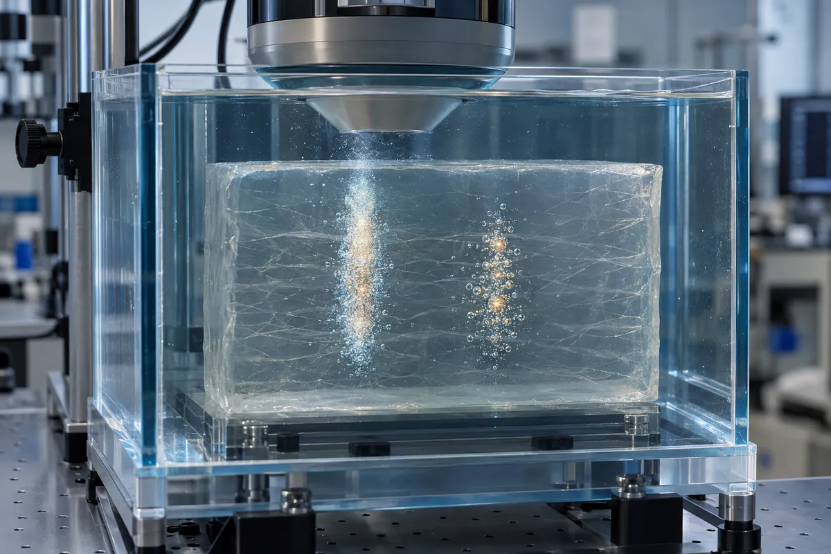
Histotripsy is a focused ultrasound technique. It is non-thermal, meaning it doesn't work by heating tissue. Instead, it relies on cavitation bubbles, tiny bubbles generated by the focused sound that mechanically break down the targeted material.
That mechanical approach is the point. The goal is to liquefy tissue at the focus without a scalpel, and without cooking it.
The surprise: same duty cycle, different outcomes
Duty cycle is the fraction of time the ultrasound is actually on. Two treatments can both run at 1% and still be built very differently: many short, rapid pulses, or fewer, longer ones separated by more downtime.
Per the Medicalxpress report, that difference mattered a lot. In tough fibrous materials, longer pulses delivered at lower repetition frequencies produced more complete liquefaction than short, rapid pulses. Total exposure was the same. The outcome was not.
Why "same dose" can mislead
It's easy to treat duty cycle like a medication dose, where the total amount is what counts. These results suggest ultrasound is more like a rhythm than a quantity. How the energy is packaged in time appears to shape what the bubbles can do to the material.
The source doesn't spell out every mechanism, so treat the following as our own reading: tough materials resist being torn apart, and pulses delivered in a different pattern may give cavitation a better chance to work against that resistance.
Why tough tissue is the hard case
Not all tissue yields equally. Fibrous tissue is built to resist mechanical stress, which is exactly the stress histotripsy applies. That is the heart of the author's original question: why do some tissues hold out?
Using tough fibrous materials as test cases is a useful way to probe the limits of the technique. If a pulse strategy can fully liquefy something resistant, it tells researchers something about what is possible in more demanding settings.
Where this could matter: BPH
Per the report, histotripsy is being explored for tougher, more fibrous tissues, including those involved in benign prostatic hyperplasia (BPH). Treatment predictability is a major challenge there.
That is where pulse design becomes practical. If timing, and not just total exposure, helps decide how completely tough tissue breaks down, then tuning pulse length and repetition frequency could be one route to more predictable noninvasive treatment. The report frames this as a possibility to be explored, not a settled clinical result.
Imaging shows different parts of the story
The account also points to the role of imaging. Different imaging methods reveal different aspects of tissue damage, so the picture of "how well did it work?" depends partly on how you look. Relying on one method could miss part of what happened to the material.
For anyone reading research in this field, that is a good habit to carry: ask not only what was measured, but how.
What to take away
When you read about an ultrasound therapy, don't stop at the headline number. Ask how the energy was delivered: pulse length, repetition rate, and not just the percentage of time the device was on. In histotripsy, that timing can be the difference between partial damage and thorough liquefaction.